Abdomen
The abdomen contains digestive organs and core muscles.

Goleisureintl · CC BY 4.0
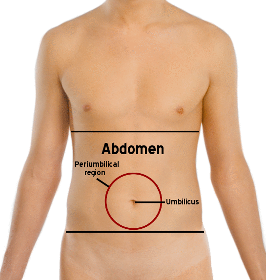
The abdomen is the front part of the torso between the thorax and pelvis in humans and other vertebrates, and the posterior tagma in arthropods. It contains most of the digestive tract and other vital organs, and its muscular wall provides protection, postural support, and assists in functions such as breathing, coughing, and childbirth.
- field
- Anatomy
- known_for
- Housing digestive and urinary organs, and providing core muscular support
Lore & Background
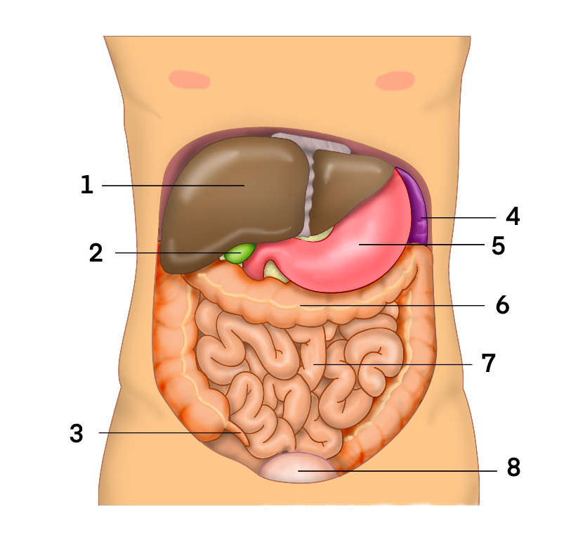
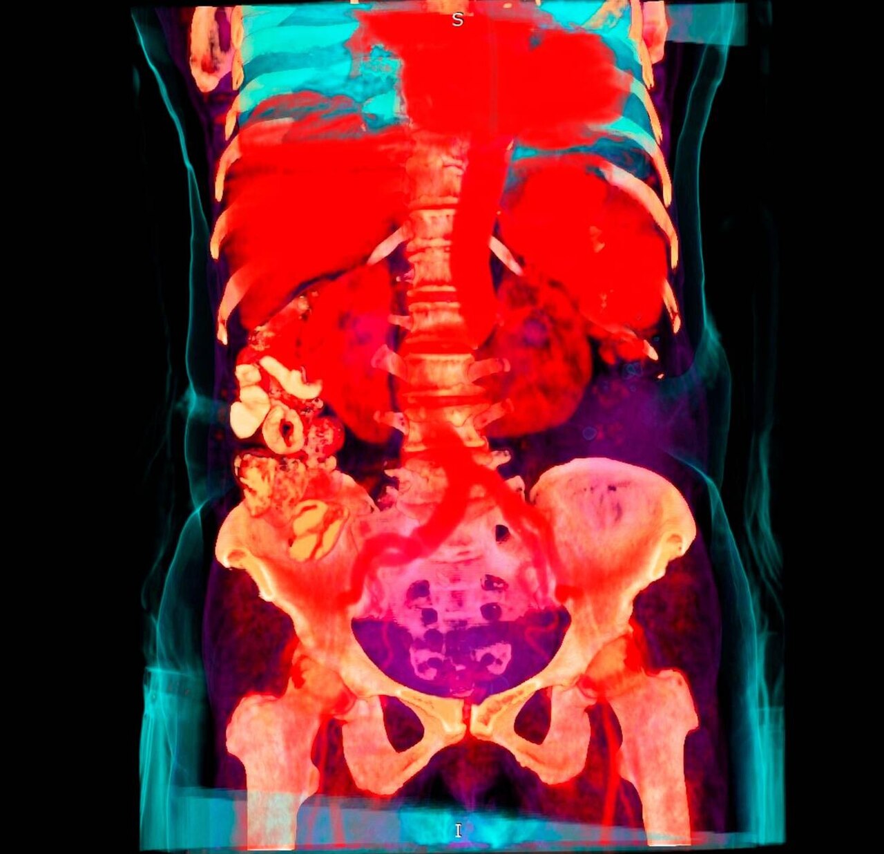
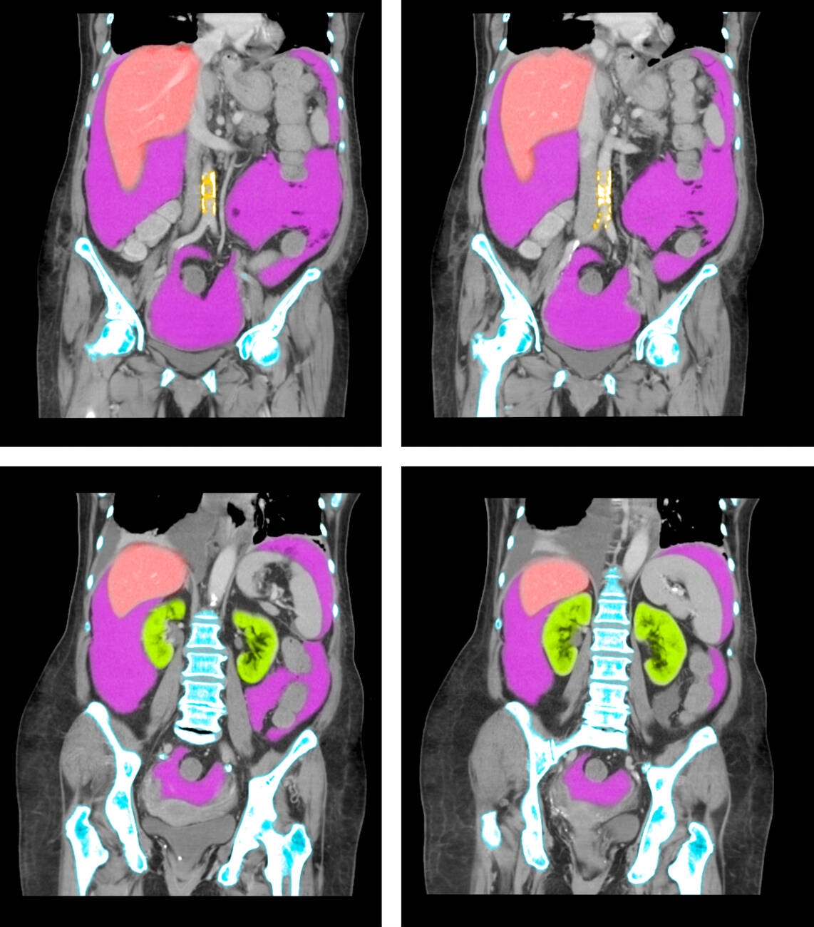
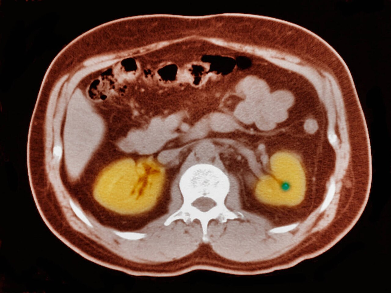
In humans, the abdomen stretches from the thoracic diaphragm to the pelvic brim, which runs from the lumbosacral joint to the pubic symphysis. The abdominal cavity is continuous with the pelvic cavity and is lined by the parietal peritoneum. Organs within the abdomen include the stomach, small intestine, colon, liver, gallbladder, pancreas, spleen, kidneys, and adrenal glands, along with major blood vessels like the aorta and inferior vena cava. The abdominal wall consists of three muscle layers: external oblique, internal oblique, and transverse abdominal, which merge toward the midline and surround the rectus abdominis. The rectus abdominis is crossed by tendinous intersections and is enclosed in a sheath formed by the lateral muscles. The pyramidalis muscle is a small triangular muscle in the lower abdomen. Functionally, the abdomen is where most digestion and absorption occur. The abdominal muscles assist in forceful exhalation, protect inner organs, provide postural support, and are involved in coughing, urination, defecation, childbirth, vomiting, and singing. They also help bend the spine forward and sideways, and assist in trunk rotation.
Reader's Guide
The abdomen is significant as the primary region for digestion and absorption of food, housing the stomach, intestines, liver, pancreas, and other organs. Its muscular wall is crucial for core stability, posture, and protection of internal organs. The three layers of abdominal muscles—external oblique, internal oblique, and transverse abdominal—work together to support the spine and enable movements such as bending and rotation. Weakness in these muscles can lead to back pain and injuries, while proper exercise improves posture and reduces the likelihood of back problems. Clinically, abdominal obesity is linked to heart disease, asthma, and type 2 diabetes, and abdominal trauma can cause severe blood loss and infection. The abdomen's social and cultural perceptions vary, with bare abdomens sometimes being sexualized and excess weight viewed differently across societies.
Did You Know?
- The abdominal cavity is continuous with the pelvic cavity and lined by the parietal peritoneum.
- The rectus abdominis muscle is crossed by three fibrous bands called tendinous intersections.
- The transverse abdominal muscle is the deepest abdominal muscle and cannot be touched from the outside.
- Abdominal obesity is associated with a higher risk of heart disease, asthma, and type 2 diabetes.
The Quadrant System and Its Diagnostic Purpose
The human abdomen is partitioned into four quadrants by anatomists and physicians to serve as a practical framework for study, diagnosis, and treatment. By dividing the abdominal area at the midline and the umbilical plane, clinicians gain a structured way to pinpoint where pain, tenderness, scars, or lumps appear, thereby narrowing down which internal organs or tissues might be implicated in a patient's symptoms. The four zones are named from the patient's own perspective: left lower quadrant, left upper quadrant, right upper quadrant, and right lower quadrant. The left lower quadrant encompasses the left iliac fossa plus half of the left flank region, while the left upper quadrant stretches from the umbilical plane up to the left ribcage. The right upper quadrant mirrors this arrangement on the patient's right side, and the right lower quadrant extends from the umbilical plane down to the right inguinal ligament. This systematic approach transforms a vague complaint into a focused anatomical investigation, guiding both physical examination and subsequent treatment decisions.
The Nine-Region Framework for Greater Precision
For more detailed anatomical mapping, the abdomen can be subdivided into nine regions using two vertical and two horizontal reference lines. The vertical lines are the mid-clavicular lines, drawn from the midpoint of each clavicle, while the upper horizontal line is the subcostal line traced along the inferior portions of the lowest costal cartilages. The lower horizontal line is the transtubercular line, connecting the tubercles of the pelvis. The three central regions running from top to bottom are the epigastric, umbilical, and hypogastric (also called pubic) region. Flanking these are six lateral regions: the left and right hypochondriac regions beside the epigastrium, the left and right lumbar flank regions beside the umbilical region, and the left and right iliac or inguinal regions beside the hypogastrium. The terminology is deliberate: "hypo" means below, "epi" means above, "chondron" refers to cartilage, and "gaster" means stomach. The left-right reversal is intentional, because anatomical designations always reflect the patient's own orientation rather than the observer's viewpoint.
Clinical Significance and Age-Specific Differential Diagnosis
The quadrant and region system is far more than an academic exercise; it directly shapes clinical reasoning at the bedside. Pain localised to the left lower quadrant may point to colitis, diverticulitis, ureteral colic, ovarian cysts, or pelvic inflammatory disease, with tumours such as colon cancer or ovarian tumours also within the differential. The left upper quadrant can signal intestinal malrotation, while the right upper quadrant raises suspicion of hepatitis, cholecystitis, or a peptic ulcer. The right lower quadrant, particularly the right inguinal region or right iliac fossa, is classically associated with appendicitis. The differential also shifts with age: children may present with gastroenteritis, intussusception, or Henoch-Schönlein purpura; adults with regional enteritis, ectopic pregnancy, or testicular torsion; and the elderly with diverticulitis, mesenteric ischaemia, or a leaking aortic aneurysm. This layered, location-based thinking ensures the full spectrum of possibilities is considered for any given site.
Comparative Anatomy and the Logic of Terminology
The quadrant nomenclature described here is specific to humans and does not carry over into comparative anatomy, because most other animals do not stand erect. In veterinary and zoological contexts, the equivalent zones are described using anterior and posterior terminology rather than upper and lower. The left lower quadrant corresponds to the left posterior quadrant in other species, the left upper quadrant to the left anterior quadrant, the right upper quadrant to the right anterior quadrant, and the right lower quadrant to the right posterior quadrant. This distinction underscores that the human system is built around an upright posture, with "upper" and "lower" referencing the vertical axis of a standing body. The anatomical designations consistently reflect the patient's own right and left, not the observer's, which is why the left and right terms appear reversed from a viewer's perspective. Grasping these conventions prevents miscommunication between human medicine and the broader field of animal anatomy.
Gallery

Frequently Asked Questions
Who is Abdomen?
The abdomen is the front section of the torso located between the thorax and the pelvis in humans and other vertebrates, while in arthropods it corresponds to the posterior tagma. It functions as the body's primary cavity for sheltering the digestive and urinary systems.
What are Abdomen's powers/role?
Its core job is to house most of the digestive tract and other vital organs while its muscular wall delivers protection, postural support, and mechanical assistance for breathing, coughing, and childbirth. Think of it as both a protective shell and a functional muscle engine.
How does Abdomen's story end?
Anatomically, the abdominal region transitions into the pelvic cavity at the pelvic brim, where the peritoneal space shifts into the pelvis. Beyond that boundary, the organs and supporting structures are no longer classified as abdominal.
Why is Abdomen important?
Without the abdominal cavity the body would have no protected space for digestion and no muscular core for posture, movement, or assisted respiration. It is the structural and metabolic hub that ties the digestive, urinary, and core-support systems together.
What's the difference between Abdomen in vertebrates vs. arthropods?
In vertebrates the abdomen is a soft-tissue region bounded by the rib cage above and the pelvis below, emphasizing internal organ housing. In arthropods it is a segmented posterior body section (the tagma) that typically carries reproductive and respiratory structures externally.
More in Animal Anatomy 1-17
Spotted an error? Know more?
This is a living reference — every entry is fact-audited, and reader corrections feed straight into our audit queue. Suggest an edit · See this site's audit record