
Cell membrane
Semipermeable lipid bilayer separating cell interior from environment.

Blackout Sea · CC0
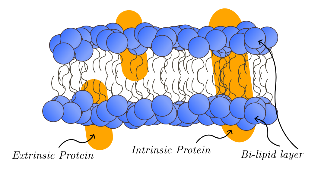
The cell membrane, also known as the plasma membrane or cytoplasmic membrane, and historically referred to as the plasmalemma, is a semipermeable biological membrane that separates and protects the interior of a cell from the outside environment. It is a lipid bilayer, usually consisting of phospholipids and glycolipids, with sterols such as cholesterol interspersed in eukaryotes and some archaea to maintain fluidity. The membrane also contains integral and peripheral proteins, and controls the movement of substances in and out of cells, being selectively permeable to ions and organic molecules. It is involved in cellular processes such as adhesion, ion conductivity, and signaling, and serves as an attachment surface for extracellular structures like the cell wall and glycocalyx, as well as the intracellular cytoskeleton.
- field
- Cell biology
- known_for
- Lipid bilayer structure, selective permeability, and role in cellular processes
- key_researchers
- Robert Hooke, Ernest Overton, Gorter and Grendel, Davson and Danielli, Singer and Nicolson
Lore & Background
Initially it was believed that all cells contained a hard cell wall since only plant cells could be observed. Microscopists focused on the cell wall for well over 150 years until advances in microscopy were made. In the early 19th century, cells were recognized as separate entities bound by individual cell walls, and this theory extended to animal cells. By the second half of the 19th century, microscopy was still not advanced enough to distinguish between cell membranes and cell walls, though some microscopists inferred cell membranes existed in animal cells due to intracellular movement.
Reader's Guide
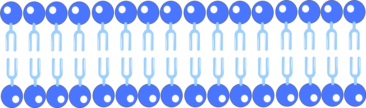
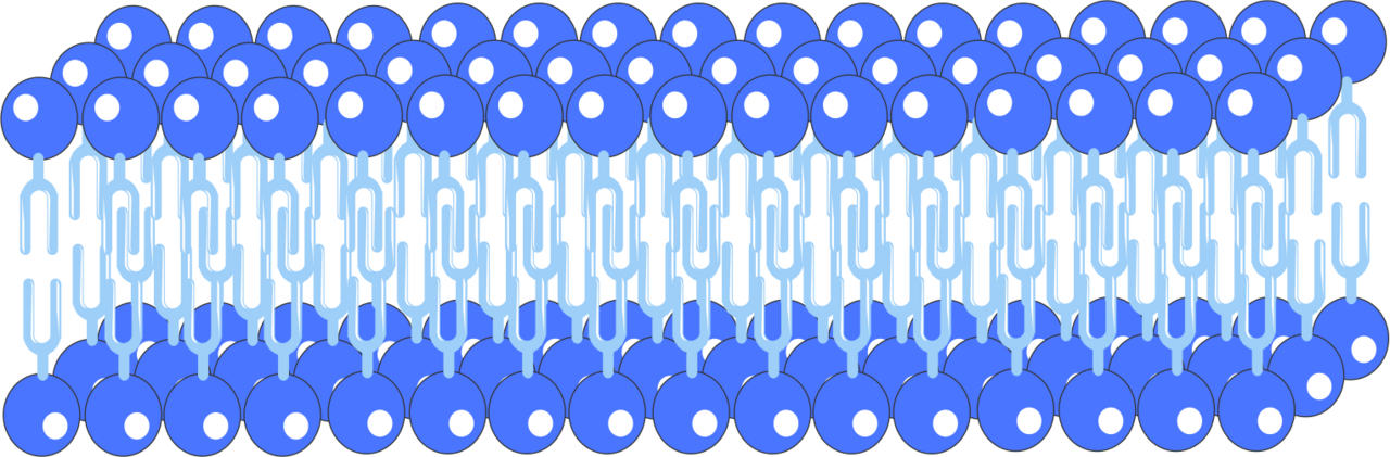
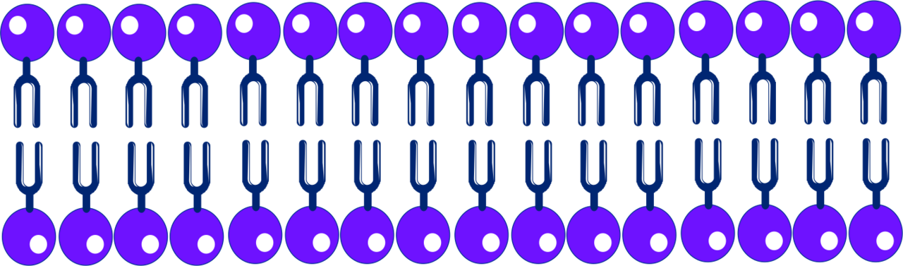
They extracted lipid from human red blood cells and measured the surface area the lipid would cover when spread on water. Since mature mammalian red blood cells lack nuclei and cytoplasmic organelles, all extracted lipids were assumed to reside in the plasma membrane. The ratio of surface area covered by extracted lipid to the surface area of the red blood cells was approximately 2:1, leading to the conclusion that the plasma membrane contains a lipid bilayer. The fluid mosaic model remains the primary archetype, describing a lipid bilayer with hydrophilic exterior heads and hydrophobic interior, where proteins interact through polar or hydrophobic interactions. The discovery by Gorter and Grendel initiated many new studies globally, confirming the structure and functions of the cell membrane are widely accepted.
The Discovery and Codification of Cell Theory
He borrowed the Latin word cellula, meaning "small room," to name what he had found, and in doing so introduced a term that would become foundational to all of biology. For nearly two centuries the concept of the cell remained loosely defined until 1839, when Matthias Jakob Schleiden and Theodor Schwann formalized cell theory. Their framework rested on three pillars: every living organism is built from one or more cells, the cell serves as the basic structural and functional unit of life, and every new cell arises from a pre-existing one. This tripartite statement unified the previously fragmented observations of botanists and zoologists into a single coherent doctrine. The theory also implicitly acknowledged that the cell is enclosed by a semipermeable membrane that holds cytoplasm and genetic material within a bounded space, establishing the membrane as the defining boundary of the living unit.
The Membrane as a Protective and Selective Barrier
At the most fundamental level, every biological cell is defined by a semipermeable membrane that encloses its cytoplasm and genetic material, separating the interior from the external environment. In bacteria this boundary is reinforced by a layered cell envelope. The plasma membrane is typically overlaid by a peptidoglycan cell wall, and in some species a third gelatinous capsule made of polysaccharides, polypeptides, or hyaluronic acid adds yet another protective shell. This architecture acts as both a mechanical and chemical filter, shielding the cell and preventing it from bursting under osmotic pressure in hypotonic conditions. Not all bacteria follow this pattern, however. Mycoplasma species possess only a cell membrane with no wall, making them uniquely vulnerable. Archaea present an even more distinctive chemistry: their membranes are built from ether-linked lipids rather than the ester-linked varieties found in bacteria and eukaryotes. The archaeon Thermoplasma, like Mycoplasma, dispenses with a cell wall entirely, relying solely on its membrane for structural integrity.
Membrane-Bound Compartmentalization and Its Exceptions
The presence or absence of internal membranes is one of the sharpest dividing lines between prokaryotic and eukaryotic cells. Prokaryotes, encompassing all bacteria and archaea, lack a membrane-bound nucleus; instead their DNA resides in a nucleoid region in direct contact with the cytoplasm. Eukaryotic cells, by contrast, enclose their genome within a nuclear membrane and house additional membrane-bound organelles such as mitochondria for energy production and chloroplasts for photosynthesis in plants. Yet the boundary is not absolute. Certain magnetotactic bacteria possess a membrane-bound organelle called the magnetosome, and anammox bacteria contain a structure known as the anammoxosome, both representing rare membrane-enclosed compartments in the prokaryotic world. Protein-based microcompartments like gas vesicles, carboxysomes, and encapsulin nanocompartments also exist in some bacteria, though they are not membrane-bound. Ribosomes, present in both domains, are membrane-less proteinaceous structures. This mosaic of compartmentalization strategies reveals that the membrane is not a binary feature but a spectrum of cellular organization.
Scale, Specialization, and the Diversity of Cellular Life
Cells first appeared on Earth roughly four billion years ago, and since then they have diversified into an extraordinary range of forms and functions. Most are visible only under a microscope, with typical prokaryotes spanning 0.1 to 5.0 micrometers in diameter. The extreme outlier is Thiomargarita magnifica, a bacterium averaging one centimeter in length and occasionally reaching two, large enough to be seen with the naked eye. Beyond size, cells vary enormously in capability. Most retain the ability to replicate and synthesize proteins, but highly differentiated types such as red blood cells and gametes have lost these functions. Some cells are motile, propelled by flagella that thread through the membrane and wall, or by the structurally distinct archaellum found in archaea. Attachment and communication are handled by pili and fimbriae, the latter built from the antigenic protein pilin. Eukaryotes range from single-celled protists, yeasts, and microalgae to the vast multicellular kingdoms of animals, plants, and fungi, each organism a consortium of many specialized cell types working in concert.
Gallery


Frequently Asked Questions
Who is Cell membrane?
The cell membrane—also called the plasma membrane or, in older literature, the plasmalemma—is the semipermeable boundary that encloses every cell and keeps its internal chemistry distinct from the surrounding environment. Structurally, it is a phospholipid bilayer studded with integral and peripheral proteins, with sterols like cholesterol interspersed in eukaryotes to tune fluidity.
What are Cell membrane's powers or role?
Its signature ability is selective permeability: it acts as a molecular gatekeeper, permitting specific ions and organic molecules to cross while blocking others. It also serves as a structural shield for the cytoplasm and as a platform for transport proteins, receptors, and cell-recognition glycolipids that drive signaling and adhesion.
How does Cell membrane's story end?
In a living cell the membrane never truly 'ends'—lipids and proteins are continuously synthesized, inserted, and recycled, so the structure is in perpetual flux. When the cell dies, the membrane loses its selective barrier function, its integrity disintegrates, and its components are eventually broken down during decomposition.
Why is Cell membrane important?
Without a defined boundary, a cell could not maintain the ion gradients, pH, and solute concentrations required for metabolism, and processes like nutrient uptake, waste excretion, and signal transduction would be impossible. It is therefore foundational to virtually every function in animal cell biology.
Who are Cell membrane's key allies (the researchers behind its discovery)?
The modern picture of the membrane was assembled across decades: Robert Hooke first noted cellular boundaries, Ernest Overton showed the barrier was lipid-based, Gorter and Grendel proposed the two-layer arrangement, Davson and Danielli added a protein coat, and Singer and Nicolson ultimately formalized the fluid-mosaic model that remains the standard description today.
More in Animal Anatomy 1-17
Spotted an error? Know more?
This is a living reference — every entry is fact-audited, and reader corrections feed straight into our audit queue. Suggest an edit · See this site's audit record