Capillary
Smallest blood vessels enabling nutrient and waste exchange.

TiffanyChan0926 · CC BY-SA 4.0
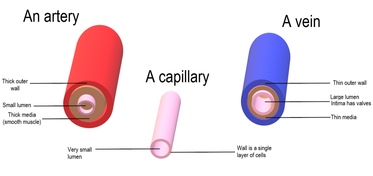
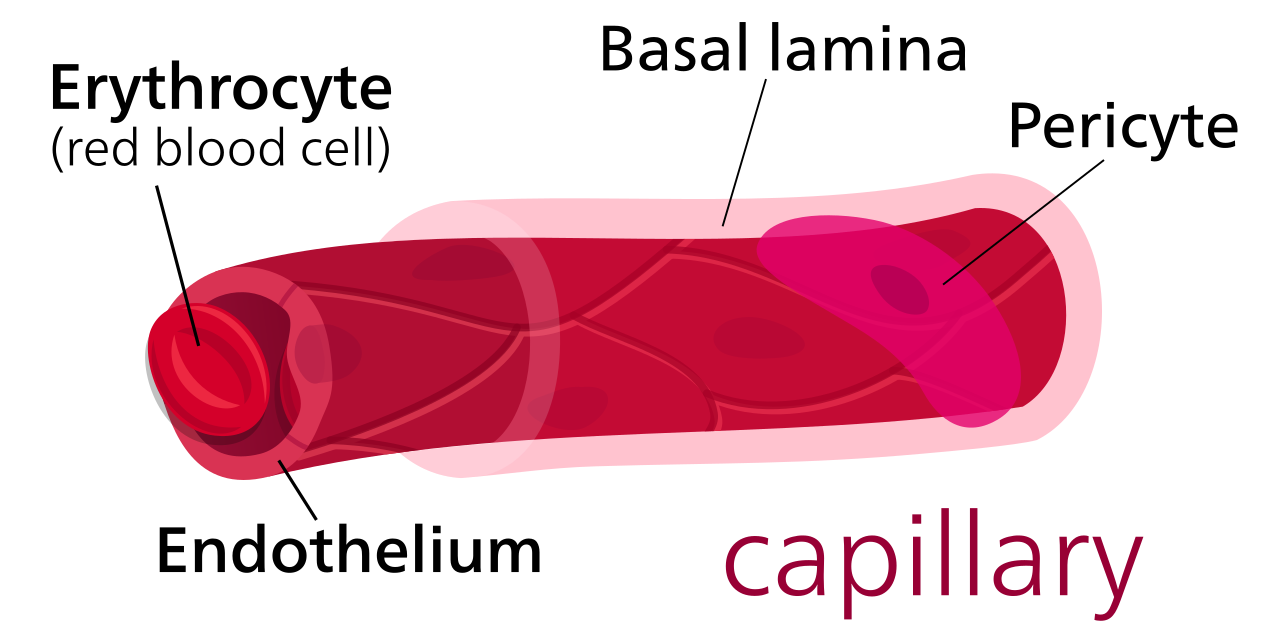
A capillary is a small blood vessel, from 5 to 10 micrometres in diameter, and is part of the microcirculation system. Capillaries are microvessels and the smallest blood vessels in the body, composed of only the tunica intima, consisting of a thin wall of simple squamous endothelial cells. They are the site of the exchange of many substances from the surrounding interstitial fluid, and they convey blood from the smallest branches of the arteries (arterioles) to those of the veins (venules).
- diameter
- 5 to 10 micrometres
- type
- Blood vessel
- part_of
- Microcirculation system
- composition
- Tunica intima of simple squamous endothelial cells
- function
- Exchange of substances between blood and interstitial fluid
- etymology
- From Latin capillaris, meaning 'of or resembling hair'
Lore & Background
Capillaries are the smallest blood vessels in the body, with a diameter of 5 to 10 micrometres. They are composed solely of the tunica intima, a thin wall of simple squamous endothelial cells. Blood flows from the heart through arteries, which branch into arterioles, and then into capillaries where nutrients and wastes are exchanged. Capillaries then join and widen to become venules, which converge into veins returning blood to the heart. Individual capillaries are part of a capillary bed, an interweaving network supplying tissues and organs; the more metabolically active a tissue, the more capillaries it requires.
Reader's Guide
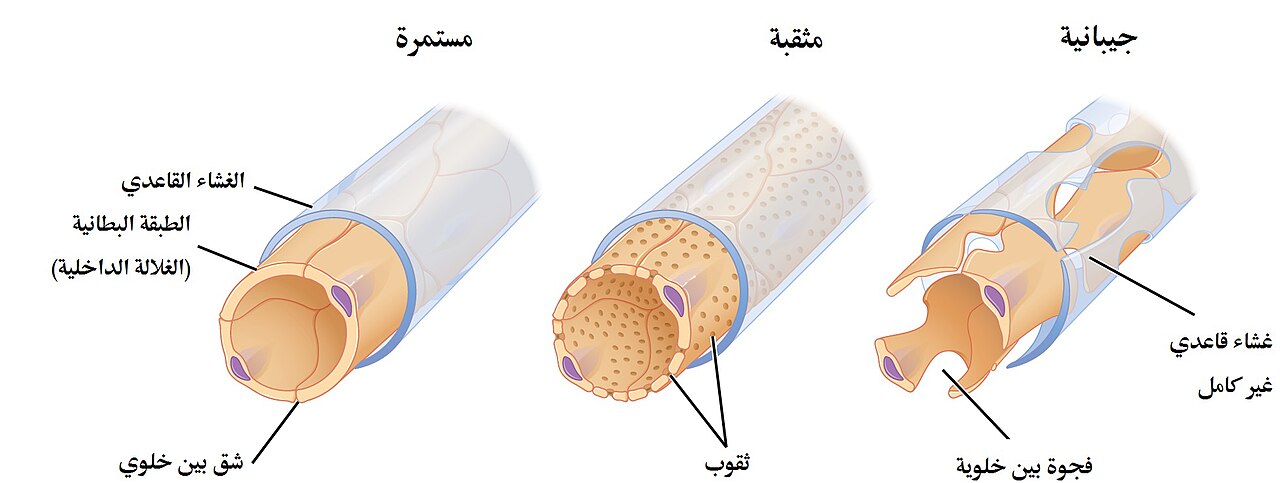
Capillaries are essential for life as they are the primary sites of exchange between blood and tissues. They allow water, oxygen, carbon dioxide, urea, glucose, and other substances to pass into and out of the interstitial fluid. Their structure varies: continuous capillaries have an uninterrupted lining and allow only small molecules through intercellular clefts; fenestrated capillaries have pores (fenestrae) that permit limited protein diffusion; sinusoidal capillaries have wider openings and allow red and white blood cells to pass. Capillary beds control blood flow via autoregulation, including myogenic response and tubuloglomerular feedback. The Starling equation quantifies the forces governing fluid exchange across capillary walls. Capillaries are also involved in the blood–brain barrier, where tight junctions prevent paracellular transport.
Did You Know?
- The word capillary comes from the Latin capillaris, meaning 'of or resembling hair', referring to their tiny diameter.
- Lymphatic capillaries are slightly larger than blood capillaries and have closed ends, allowing interstitial fluid to flow in but not out.
- Sinusoidal capillaries lack diaphragms over their pores and can allow red and white blood cells to pass through.
- Capillary permeability can be increased by cytokines, anaphylatoxins, histamine, and other immune mediators.
Anatomy of the Smallest Vessel
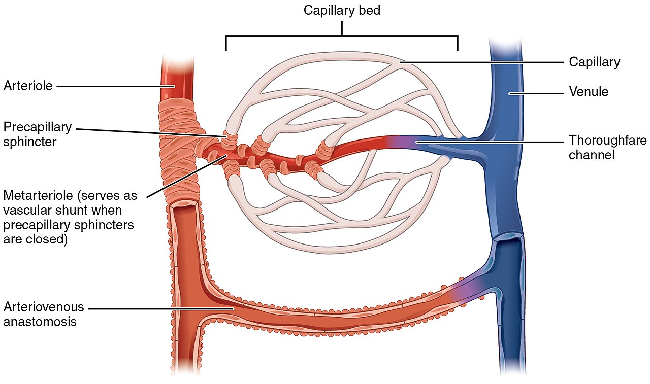
A capillary is the tiniest conduit in the human circulatory system, measuring just five to ten micrometres across. Unlike larger arteries and veins that possess multiple tissue layers, a capillary wall consists of nothing more than the tunica intima—a single, delicate sheet of simple squamous endothelial cells. This extreme thinness is no accident; it is precisely what makes the rapid exchange of oxygen, carbon dioxide, glucose, urea, and countless other solutes possible between the blood and the surrounding interstitial fluid. Capillaries do not exist in isolation. They weave together into dense, interlacing networks called capillary beds that blanket every tissue and organ. The metabolic demands of a given region dictate how densely those beds are packed: highly active tissues command far more capillary supply than quiescent ones. At one end, a capillary receives blood from an arteriole; at the other, it drains into a venule, completing the microcirculatory loop. In the mesentery, an intermediate vessel called a metarteriole adds an extra stage between arterioles and the capillary network.
Three Architectures, Three Purposes
Blood capillaries are not a single uniform design. They fall into three distinct structural categories, each tailored to the exchange needs of its host tissue. Continuous capillaries present an unbroken endothelial lining; only small molecules like water and ions slip through the narrow intercellular clefts, while lipid-soluble substances diffuse across the cell membranes themselves. Two subtypes exist: one rich in transport vesicles, found in skeletal muscle, fingers, gonads, and skin, and another with few vesicles that forms the blood–brain barrier in the central nervous system. Fenestrated capillaries take permeability a step further, puncturing their endothelium with 60-to-80-nanometre pores called fenestrae, each bridged by a fine diaphragm of radially arranged fibrils. These appear in endocrine glands, the intestines, the pancreas, and the kidney glomeruli, where podocyte foot processes add an additional filtration layer. Sinusoidal capillaries, sometimes called discontinuous, are the most open of the three. Their fenestrations span 30 to 40 micrometres, they lack any diaphragm, and their basal lamina is patchy rather than continuous. This permissive architecture lets whole red and white blood cells, along with large serum proteins, cross the wall. They are characteristic of the liver, bone marrow, spleen, and certain brain circumventricular organs.
The Exchange That Keeps Us Alive
The entire raison d'être of a capillary is to serve as a permeable interface between blood and tissue. Molecules smaller than roughly three nanometres—water, oxygen, carbon dioxide—cross the wall by squeezing through the gaps between adjacent endothelial cells, a route called paracellular transport. Larger solutes such as albumin and other big proteins cannot fit through those clefts; instead they are shuttled across the cell itself inside membrane-bound vesicles, a mechanism known as transcellular transport. Both routes operate bidirectionally, driven by osmotic and concentration gradients, so that nutrients flow into tissue while metabolic wastes like urea, lactic acid, uric acid, and creatinine flow back into the bloodstream. In the brain, however, the rules tighten dramatically. The continuous capillaries of the blood–brain barrier are sealed by tight junctions that eliminate the paracellular route entirely, leaving only carefully regulated transcellular pathways. This selective restriction is what protects neural tissue from the chemical fluctuations of the rest of the body.
From Latin Roots to Embryonic Origins
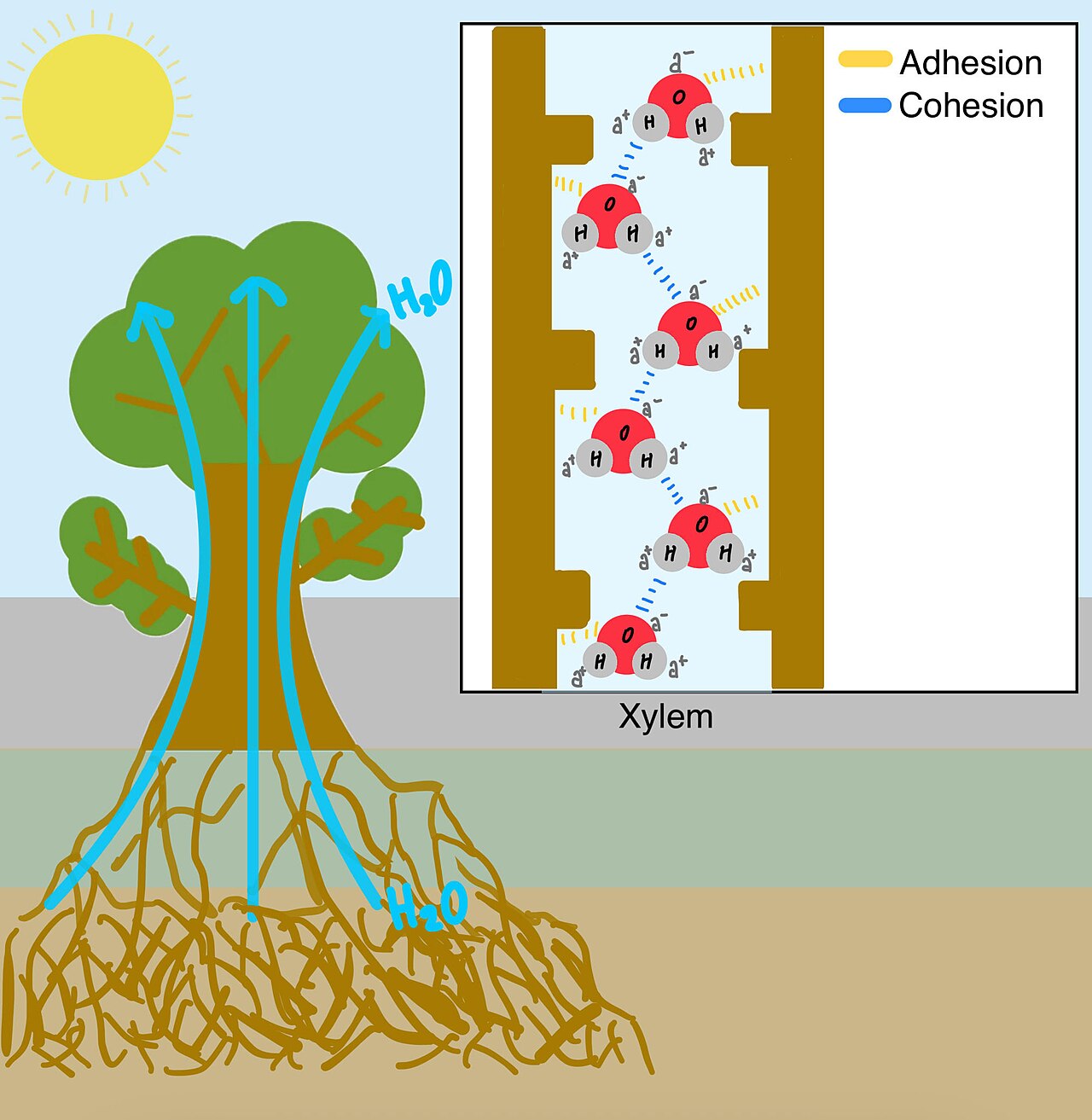
The very name "capillary" is a nod to the vessel's hairlike slenderness. It derives from the Latin capillaris, meaning "of or resembling hair," and entered English usage in the mid-seventeenth century. The word also lives on as an adjective in the phrase "capillary action," describing the way a liquid climbs through narrow spaces without the aid of gravity or other external forces. Beyond blood, the capillary concept extends into the lymphatic system. Lymphatic capillaries are slightly wider than their blood counterparts and terminate in closed ends, a structural feature that allows interstitial fluid to flow in but never out. Because lymph carries a higher concentration of plasma proteins, these vessels maintain a greater internal oncotic pressure than blood capillaries, helping to pull fluid from the tissues. The capillary network does not simply appear fully formed. During early embryonic development, vasculogenesis generates brand-new endothelial cells that assemble into primitive vascular tubes, while angiogenesis sprouts additional capillaries from already-existing vessels. Together these processes lay down the first interconnecting networks that vascularise the yolk sac, the connecting stalk, and the chorionic villi, setting the stage for the vast microcirculatory web that sustains adult life.
Gallery
Frequently Asked Questions
What is a capillary in animal anatomy?
A capillary is the smallest blood vessel in the body, measuring only 5 to 10 micrometres in diameter. It sits within the microcirculation system and acts as the final connecting link between arterioles and venules.
What is a capillary made of structurally?
Unlike larger vessels that have multiple tissue layers, a capillary wall is composed solely of the tunica intima. That means it is built from just one thin sheet of simple squamous endothelial cells.
What is the main function of a capillary?
Capillaries are the site where nutrients, oxygen, and metabolic waste are exchanged between the blood and the surrounding interstitial fluid. Their single-cell-thick wall is precisely what allows this rapid transfer to occur.
Where does a capillary sit in the blood-flow pathway?
Blood enters capillaries from the smallest arterial branches (arterioles) and drains out into the smallest venous branches (venules). In this way, capillaries serve as the bridging segment that links the arterial and venous sides of the microcirculation.
Why is it called a 'capillary'?
The name derives from the Latin word capillaris, meaning 'of or resembling hair.' The term reflects the fine, hair-like appearance these tiny vessels take when viewed under magnification.
More in Animal Anatomy 1-17
Spotted an error? Know more?
This is a living reference — every entry is fact-audited, and reader corrections feed straight into our audit queue. Suggest an edit · See this site's audit record