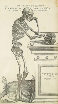
Artery
Blood vessel carrying blood away from the heart.

Wikimedia Commons · CC BY 4.0
An artery is a blood vessel in humans and most other animals that carries oxygenated blood away from the heart in the systemic circulation to one or more parts of the body. Exceptions include the pulmonary arteries, which carry deoxygenated blood to the lungs for oxygenation, and the umbilical arteries in fetal circulation, which carry deoxygenated blood to the placenta. Arteries consist of a multi-layered wall wrapped into a tube-shaped channel and contrast with veins, which carry deoxygenated blood back toward the heart.
- type
- Blood vessel
- function
- Carries blood away from the heart
- key_exceptions
- Pulmonary arteries and umbilical arteries
- wall_layers
- Tunica intima, tunica media, tunica externa
- pressure
- Higher than other parts of the circulatory system
- primary_determinants
- Cardiac output and systemic vascular resistance
Lore & Background
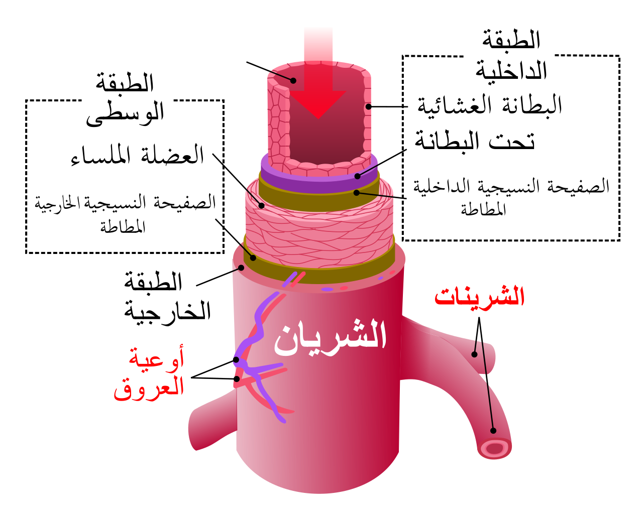
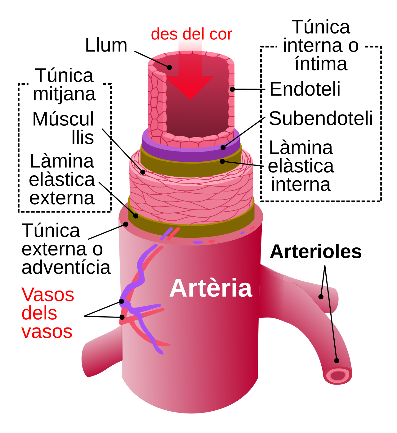
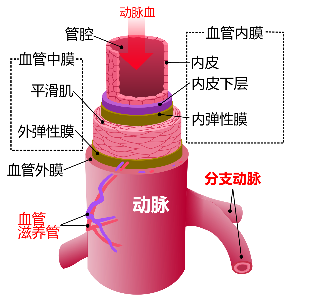
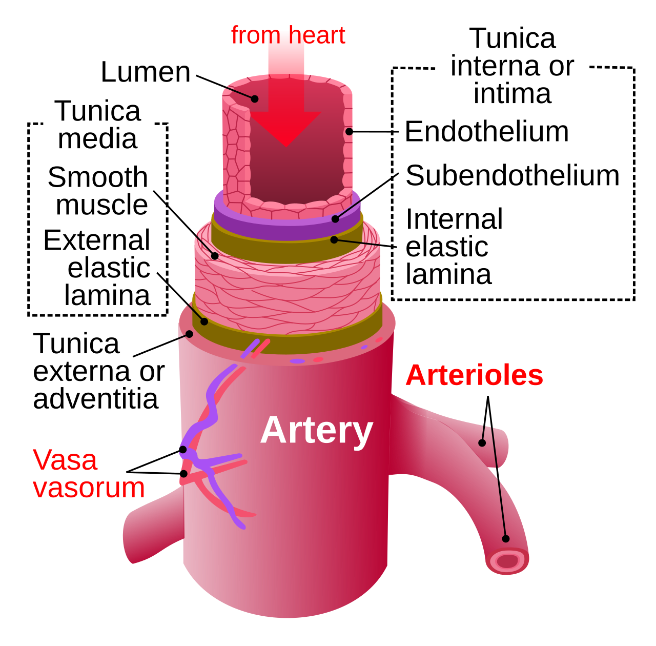
The anatomy of arteries can be separated into gross anatomy and microanatomy. The arterial system is divided into systemic arteries, carrying blood from the heart to the whole body, and pulmonary arteries, carrying deoxygenated blood from the heart to the lungs. Large arteries such as the aorta are composed of endothelial, smooth muscle, fibroblast, and immune cells. The arterial wall consists of three layers: the tunica intima, tunica media, and tunica externa, from innermost to outermost. The hollow internal cavity in which blood flows is called the lumen.
Reader's Guide
Arteries form part of the circulatory system and carry oxygenated blood away from the heart to the tissues, except for pulmonary arteries. They have a blood pressure higher than other parts of the circulatory system, varying during the cardiac cycle. The combination of cardiac output and systemic vascular resistance are the principal determinants of arterial blood pressure. Systemic arteries can be subdivided into elastic and muscular types. Arterioles have the greatest collective influence on local blood flow and overall blood pressure. Over time, factors such as elevated blood sugar, cholesterol, high blood pressure, stress, and smoking can damage artery walls, resulting in atherosclerosis. Accidental intra-arterial injection can cause intense pain, paresthesia, and necrosis, often requiring amputation.
Did You Know?
- The word 'artery' comes from Greek ἀρτηρία (artēríā).
- Pulmonary arteries carry deoxygenated blood to the lungs, unlike most arteries.
- The hollow internal cavity of an artery is called the lumen.
- Arterioles are the primary 'adjustable nozzles' in the blood system.
Anatomical Origin and Pelvic Journey
The internal pudendal artery represents the final branch emerging from the anterior trunk of the internal iliac artery, making it a terminal vessel within that arterial system. Its course through the pelvis is remarkably circuitous. After arising from the anterior division of the internal iliac, the vessel courses along the lateral pelvic wall before making its way out of the pelvic cavity entirely. It passes through the greater sciatic foramen, specifically inferior to the piriformis muscle, which deposits it into the gluteal region. From there, it hooks around the sacrospinous ligament and re-enters the pelvis via the lesser sciatic foramen to reach the perineum. Once in the perineal space, it travels alongside the internal pudendal veins and the pudendal nerve within the pudendal canal. Notably, this artery is measurably smaller in females than in males, reflecting differences in the vascular demands of the external genitalia between the sexes.
Branching Pattern and Sexual Dimorphism
The internal pudendal artery fans out into several named branches that serve the external genitalia. In females, it gives rise to the deep artery of the clitoris, which supplies the clitoral crura, as well as the dorsal artery of the clitoris. The urethral artery presents a point of anatomical debate: some authorities classify it as a direct branch of the internal pudendal artery, while others assign it to the perineal artery instead. In males, the branching pattern includes the perforating arteries of the penis, which are unique to the male perineum. The overall caliber of the artery differs between the sexes, being smaller in females. This sexual dimorphism in size and branching reflects the distinct vascular architecture required by the male and female external genital structures, and it carries practical implications for surgical and obstetric procedures where these vessels must be identified and preserved.
Clinical Relevance in Obstetrics
The most significant clinical encounter with the internal pudendal artery occurs in the context of childbirth. During vaginal delivery, the mechanical stress and stretching of the perineal tissues can compromise this vessel in women, leading to vascular injury. The most common consequence is the formation of a haematoma, a collection of blood in the surrounding soft tissues. In the majority of cases, such a haematoma is self-limiting and resolves on its own without the need for surgical intervention or other treatment. However, in a subset of cases, the pooled blood can become a medium for bacterial growth, transforming the haematoma into an infected abscess that requires more aggressive management. This clinical scenario underscores the vulnerability of the pudendal vasculature during obstetric events and highlights the importance of monitoring postpartum patients for signs of perineal swelling, pain, or systemic infection that might indicate a complication arising from injury to the internal pudendal artery or its branches.
Anatomical Variation in Males
One of the most striking features of the internal pudendal artery is its considerable anatomical variability, particularly in men. Approximately seventy percent of males possess an accessory internal pudendal artery, a finding that makes this variation the norm rather than the exception in the male population. What distinguishes this accessory vessel is its atypical origin: rather than arising from the internal iliac artery as the standard internal pudendal artery does, the accessory branch typically originates from one of several alternative sources. These include the external iliac artery, the obturator artery, or the vesical arteries. This variation carries important implications for any surgical procedure in the pelvic or perineal region, as a surgeon who is unaware of the accessory vessel's presence and its non-standard origin risks inadvertent injury. The high prevalence of this variant also means that anatomical descriptions presenting only the single, standard internal pudendal artery offer an incomplete picture of the typical male pelvic vasculature.
Gallery
Frequently Asked Questions
What is an artery in the Animal Anatomy series?
An artery is a tube-shaped blood vessel whose core job is to move blood away from the heart and out to the rest of the body. In most animals, including humans, the blood it carries is oxygen-rich, making it the delivery arm of the systemic circulation.
How does an artery differ from a vein?
Arteries handle the outbound trip, pushing blood away from the heart, whereas veins manage the return journey, bringing deoxygenated blood back. Arteries also operate under noticeably higher pressure than any other segment of the circulatory system.
Do all arteries carry oxygenated blood?
Not always. The pulmonary arteries are the classic exception, shuttling deoxygenated blood from the heart to the lungs for gas exchange, and fetal umbilical arteries likewise carry deoxygenated blood — but to the placenta rather than the lungs.
What are the three wall layers of an artery?
The arterial wall is built from three concentric layers: the innermost tunica intima, the middle tunica media, and the outermost tunica externa. Together these layers create a durable channel capable of withstanding the high-pressure flow that defines arterial function.
What determines the pressure inside an artery?
Arterial pressure is set primarily by two factors: the volume of blood the heart ejects (cardiac output) and the total resistance the blood encounters as it travels through the systemic vasculature. This combination is why arteries experience the highest pressures anywhere in the circulatory system.
More in Animal Anatomy 1-17
Spotted an error? Know more?
This is a living reference — every entry is fact-audited, and reader corrections feed straight into our audit queue. Suggest an edit · See this site's audit record