
Adrenal gland
Endocrine glands producing hormones for stress, metabolism, and salt balance.

PINSupport · CC BY-SA 4.0
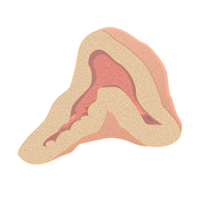
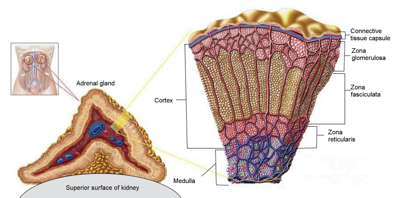
The adrenal glands (also known as suprarenal glands) are endocrine glands located above the kidneys in the retroperitoneum. They produce a variety of hormones essential for regulating metabolism, blood pressure, electrolyte balance, and stress responses, including adrenaline, aldosterone, and cortisol. Each gland consists of an outer cortex, which produces steroid hormones, and an inner medulla, which produces catecholamines.
- location
- Above and slightly medial to the kidneys, in the retroperitoneum
- shape
- Right pyramidal; left semilunar or crescent shaped and somewhat larger
- size
- Approximately 5 cm in length, 3 cm in width, up to 1 cm in thickness
- weight
- Combined weight 7 to 10 grams in an adult human
- color
- Yellowish
- blood supply
- Up to 60 small arteries per gland; three main arteries supply each gland
Lore & Background
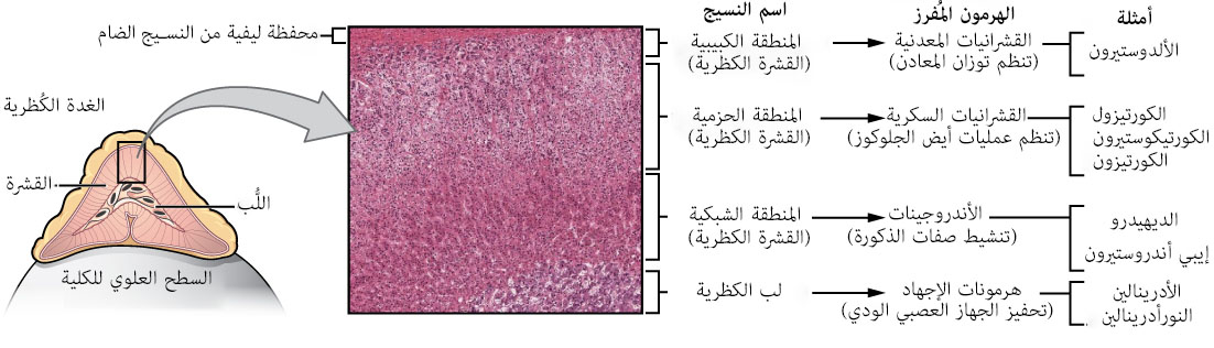
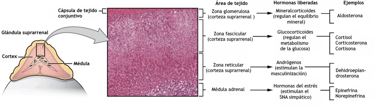
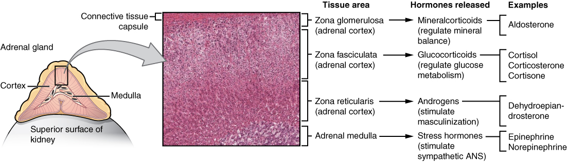
The adrenal glands are divided into two distinct parts: the outer adrenal cortex and the inner medulla. The cortex itself has three zones: the zona glomerulosa produces mineralocorticoids like aldosterone, which regulate blood pressure and electrolyte balance; the zona fasciculata produces glucocorticoids such as cortisol, which regulate metabolism and suppress the immune system; and the zona reticularis produces androgens that are converted to sex hormones elsewhere. The medulla produces catecholamines—adrenaline and noradrenaline—which enable rapid stress responses.
Reader's Guide
The adrenal glands are critical for homeostasis and survival, producing hormones that influence nearly every organ system. Their dysfunction leads to significant endocrine diseases: overproduction of cortisol causes Cushing's syndrome, while insufficient production leads to Addison's disease. Congenital adrenal hyperplasia results from genetic dysregulation of endocrine control. Tumors arising from adrenal tissue are commonly found incidentally during medical imaging. The glands' rich blood supply—up to 60 small arteries per gland—underscores their metabolic importance. Their unique structure, including the longitudinally oriented smooth muscle bundles in the central adrenomedullary vein, reflects specialized functions. Understanding adrenal physiology is essential for diagnosing and treating disorders of blood pressure, metabolism, and stress response.
Did You Know?
- The adrenal glands have one of the greatest blood supply rates per gram of tissue of any organ.
- The adrenal medulla can be considered a specialized sympathetic ganglion because it is innervated by preganglionic nerve fibers.
- The zona fasciculata accounts for nearly 80% of the volume of the adrenal cortex.
- The right adrenal gland is pyramidal in shape, while the left is semilunar or crescent shaped and somewhat larger.
Anatomical Home in the Abdominal Landscape
The adrenal glands — a pair of small organs — find their place within the broader abdominal and pelvic region of the human body. In the layered architecture of anatomy, they sit at the organ level: the body is built from biological systems, which in turn are composed of organs, which are made of tissues, which are constructed from cells and connective tissue. This hierarchical nesting means that understanding the adrenal glands requires grasping both their position among neighboring structures (kidneys, liver, pancreas, spleen, stomach, intestines, bladder, gallbladder, and appendix) and their internal tissue organization. Gross anatomy, the study of structures visible to the naked eye, places the adrenal glands within the abdominal and pelvic map. Microscopic anatomy, examined through histology and cytology with the aid of microscopes, reveals the finer tissue and cellular architecture that defines their function. Together, these two levels of study give a complete picture of where these paired glands reside and how they are built.
A Voice in the Endocrine Chorus
The adrenal glands belong to the endocrine system, a major organ system whose defining purpose is internal communication through hormones produced by endocrine glands. In this hormonal dialogue, the adrenal glands are listed alongside the hypothalamus, pituitary gland, pineal gland, thyroid, and parathyroid glands — a constellation of signaling organs that together coordinate the body's internal messaging. The endocrine system operates as one of the body's principal biological systems, sitting alongside the circulatory, digestive, immune, integumentary, lymphatic, musculoskeletal, nervous, reproductive, respiratory, and urinary systems. Understanding the adrenal glands in this context means recognizing them not as isolated structures but as participants in a body-wide chemical conversation. Anatomy, physiology, and biochemistry — the three complementary basic medical sciences — work in tandem to explain not just where the adrenal glands sit, but how they function and what chemistry underlies their hormonal output.
Tracing the Adrenal Gland Through History
The long history of human anatomy has been marked by a steadily deepening comprehension of what organs actually do, and the adrenal glands are no exception to this evolving understanding. Early anatomical investigation relied on examining animals and dissecting fresh or preserved cadavers, methods that gradually gave way to the technologically complex techniques developed during the twentieth century. Each era brought new tools and perspectives: where once a surgeon's hands and a scalpel revealed the shape and position of abdominal organs, later generations could examine the minute tissue and cellular structures of the adrenal glands through histological preparations viewed under a microscope. The progression from gross anatomical observation to microscopic cytology and histology transformed how these paired glands were understood. For students and practitioners, this historical arc means the adrenal gland is known today through a layered accumulation of methods — from naked-eye identification of its position in the abdomen and pelvis to detailed cellular analysis revealing its internal organization.
Teaching the Adrenal Gland to a New Generation
Learning about the adrenal glands is a standard component of medical and biological education. Physicians, dentists, physiotherapists, nurses, paramedics, radiographers, and students in certain biological sciences encounter them through anatomical models, skeletons, textbooks, diagrams, photographs, lectures, and tutorials. Medical and dental students additionally gain hands-on experience through dissection and inspection of cadavers, and they examine histological slides under a microscope to study the gland's tissue architecture. In the classroom, the adrenal glands can be approached regionally — as part of the abdomen and pelvis unit — or systemically — as members of the endocrine system. The landmark textbook Gray's Anatomy was recently reorganized from a systems-based format to a regional one, aligning with modern teaching preferences. A thorough working knowledge of anatomy, including the adrenal glands, is required of all medical doctors, with particular importance for surgeons and those in diagnostic specialities such as histopathology and radiology.
Gallery

Frequently Asked Questions
Who is Adrenal gland?
The adrenal gland (also called the suprarenal gland) is a pair of small endocrine organs that sit perched on top of each kidney in the retroperitoneal space. The right one is pyramidal while the left is crescent-shaped and slightly larger, and together they weigh only about 7 to 10 grams in an adult human.
What are Adrenal gland's powers/role?
Each gland has two functional zones: an outer cortex that synthesizes steroid hormones such as cortisol and aldosterone, and an inner medulla that releases catecholamines like adrenaline. Together these secretions govern the body's acute stress response, metabolic rate, blood pressure, and electrolyte balance.
Where does Adrenal gland live?
The pair resides directly above and slightly medial to the kidneys, tucked into the retroperitoneum. Blood reaches them through up to sixty tiny arteries per gland, with three main arterial branches serving as the primary supply lines.
How does Adrenal gland's story end?
If both glands are removed or destroyed, the organism cannot survive without exogenous hormone replacement because cortisol and aldosterone have no adequate backup source. A single remaining gland, however, can usually compensate for the other's absence and keep the individual alive.
Why is Adrenal gland important?
Without adrenal output, the body loses its primary chemical toolkit for handling acute stress, maintaining sodium-potassium balance, and keeping blood pressure stable. That makes even these two tiny yellowish organs—roughly 5 cm long, 3 cm wide, and up to 1 cm thick—absolutely critical to everyday survival.
More in Animal Anatomy 1-17
Spotted an error? Know more?
This is a living reference — every entry is fact-audited, and reader corrections feed straight into our audit queue. Suggest an edit · See this site's audit record