Brainstem
Posterior brain part connecting cerebrum to spinal cord.

DataBase Center for Life Science (DBCLS) · CC BY 4.0
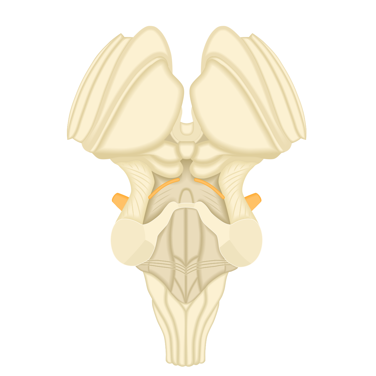
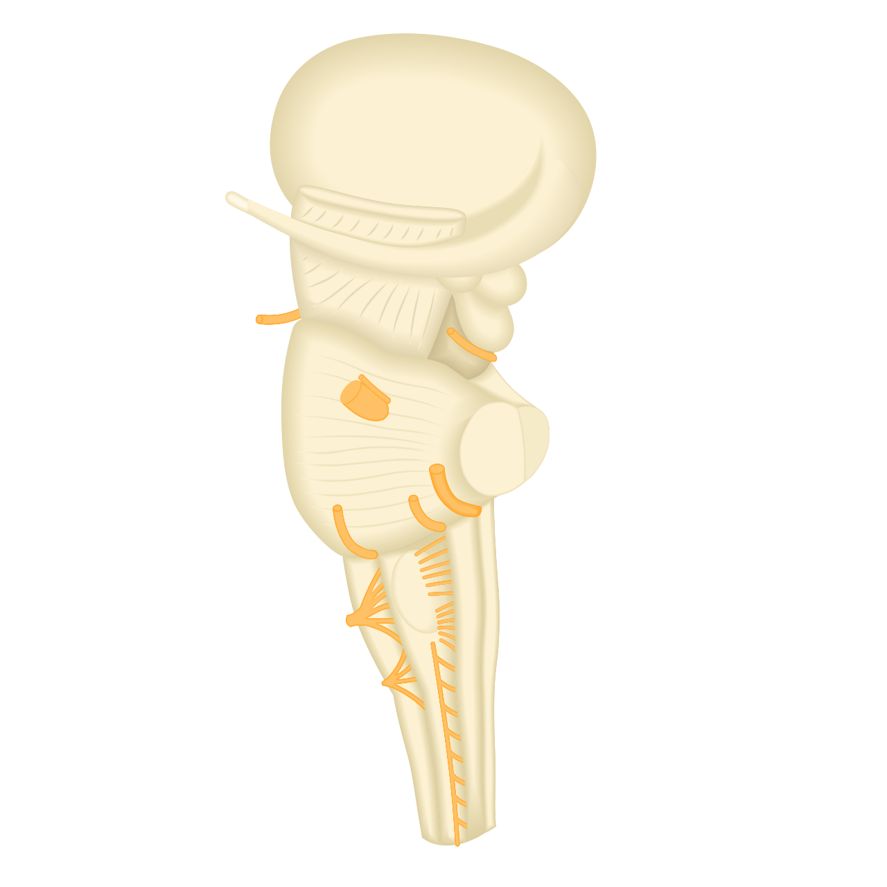
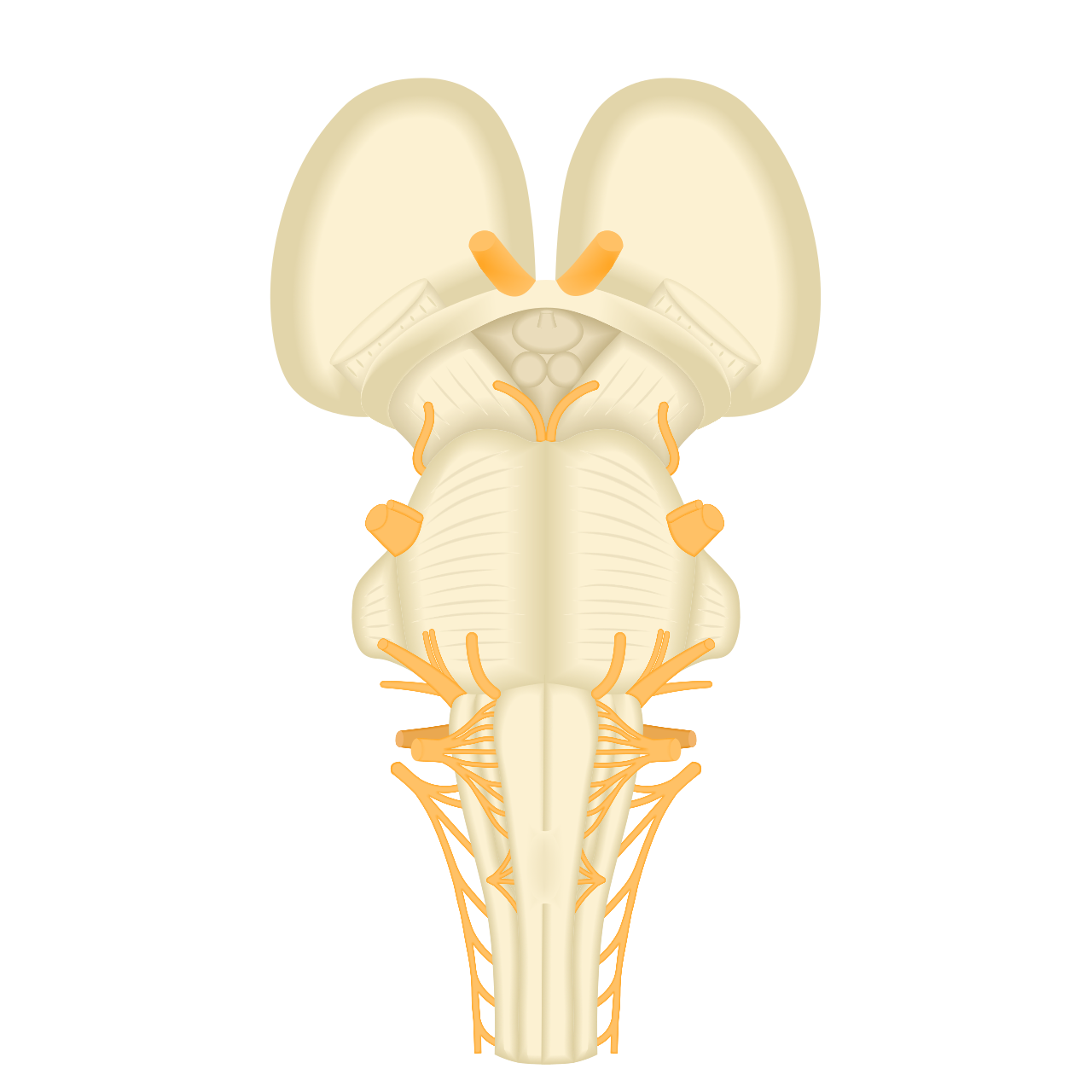
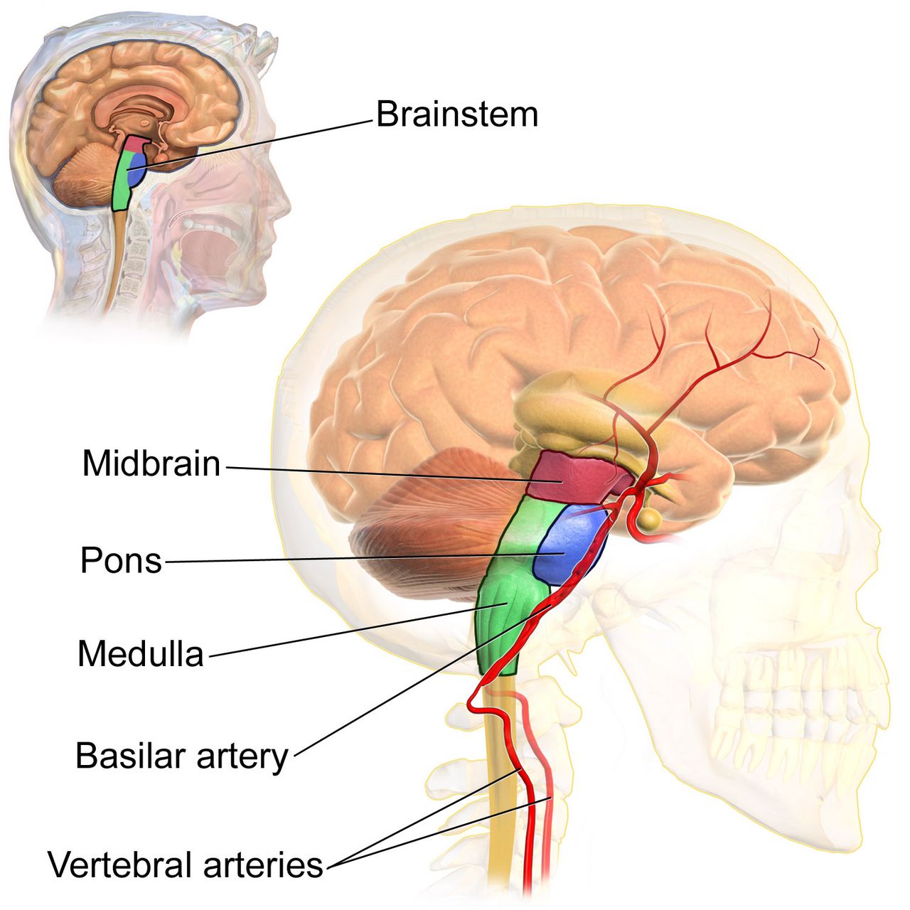
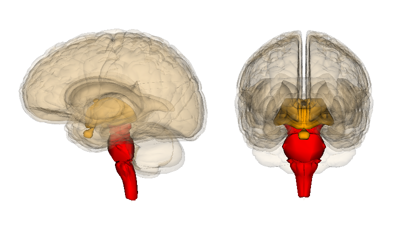
The brainstem is the posterior stalk-like part of the brain that connects the cerebrum with the spinal cord. In the human brain, it is composed of the midbrain, the pons, and the medulla oblongata, with the diencephalon sometimes included. Despite making up only about 2.6 percent of the brain's total weight, it has critical roles in regulating heart and respiratory function, controlling heart rate and breathing rate, and providing the main motor and sensory nerve supply to the face and neck via the cranial nerves.
- composition
- Midbrain, pons, medulla oblongata (diencephalon sometimes included)
- weight_percentage
- 2.6% of brain's total weight
- cranial_nerves_originating
- Ten pairs
- key_functions
- Regulating heart rate, breathing rate, sleep cycle, motor and sensory pathways
- location
- Extends from just above tentorial notch to first cervical vertebra below foramen magnum
Lore & Background
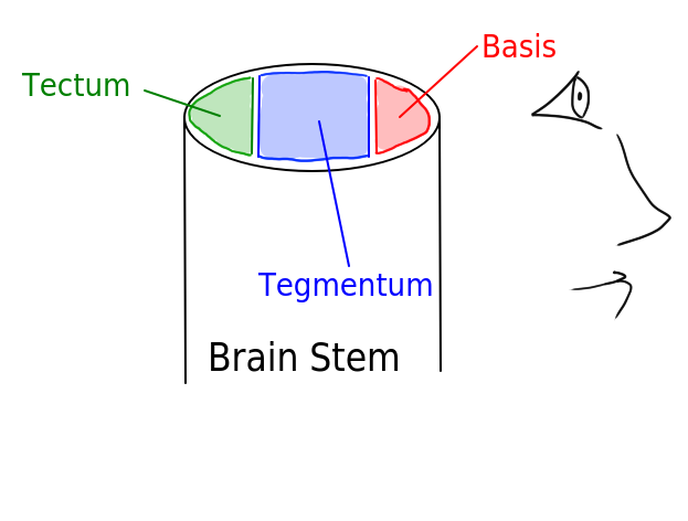
The brainstem is composed of three main parts: the midbrain, pons, and medulla oblongata. The midbrain is further subdivided into the tectum, tegmentum, and ventral tegmental area. The tectum comprises the superior and inferior colliculi, involved in vision and audition respectively. The tegmentum contains several nuclei, tracts, and the reticular formation. The ventral tegmental area includes paired cerebral peduncles that transmit axons of upper motor neurons. The pons lies between the midbrain and medulla, containing tracts that carry signals between the cerebrum, medulla, and cerebellum, and houses respiratory centers. The medulla oblongata is continuous with the spinal cord and contains cardiac, respiratory, and vasomotor centers.
Reader's Guide
The brainstem is of prime importance in conveying motor and sensory pathways from the rest of the brain to the body and back. These pathways include the corticospinal tract for motor function, the dorsal column-medial lemniscus pathway for fine touch and proprioception, and the spinothalamic tract for pain and temperature. It also regulates the central nervous system and the body's sleep cycle. Ten pairs of cranial nerves originate from the brainstem, supplying motor and sensory functions to the face and neck. Key nuclei within the midbrain include the periaqueductal gray involved in pain desensitization, the red nucleus for motor function, and the substantia nigra pars compacta, whose dysfunction is implicated in Parkinson's disease. The pons coordinates activities of the cerebellar hemispheres, and the medulla controls vomiting via the area postrema. The brainstem's small size belies its critical role in sustaining life.
Did You Know?
- The brainstem makes up only about 2.6 percent of the brain's total weight.
- Ten pairs of cranial nerves come from the brainstem.
- The substantia nigra pars compacta in the midbrain uses dopamine as its neurotransmitter and its dysfunction is implicated in Parkinson's disease.
- The medulla oblongata contains the area postrema, which controls vomiting.
Architecture & Spatial Organization
The reticular formation defies easy anatomical boundaries. Rather than forming a single compact mass, it constitutes a diffuse, net-like web of roughly one hundred nuclei scattered throughout the core of the brainstem, stretching from the lower medulla oblongata to the upper midbrain. In practical terms, it can be understood as the collection of interspersed cells filling the spaces between the more clearly defined, named structures. This diffuse architecture is nonetheless organized. Traditionally, the reticular nuclei are sorted into three sagittal columns. The median column houses the raphe nuclei, which form a central ridge. Flanking this ridge on the medial side are the gigantocellular nuclei, named for their relatively large cell bodies, while the lateral column contains the smaller parvocellular nuclei. The medial reticular formation is notably large and carries long ascending and descending fibers, whereas the lateral reticular formation sits close to the motor nuclei of the cranial nerves and largely mediates their reflexive functions. As one traces the structure caudally from the rostral midbrain, the medial component fades in prominence while the lateral component grows more conspicuous, particularly in the rostral medulla and caudal pons, where major cranial nerves including the vagus take their origin.
The ARAS & the Chemistry of Wakefulness
Perhaps no single subsystem of the reticular formation has captured more scientific attention than the ascending reticular activating system, often described as a biological on-off switch for the cerebral cortex. Located primarily within the midbrain reticular formation, the ARAS is a sprawling collection of more than twenty nuclei on each side, distributed across the upper brainstem, pons, medulla, and posterior hypothalamus. Its power lies in chemical diversity. The neurons of the ARAS release an impressive array of neurotransmitters—dopamine, norepinephrine, serotonin, histamine, acetylcholine, and glutamate—each contributing to the regulation of wakefulness and sleep-wake transitions. These signals reach the cortex through two principal routes: direct axonal projections and indirect pathways relayed through the thalamus. The thalamic route is dominated by cholinergic neurons in the pontine tegmentum, while the hypothalamic route employs a different neurochemical profile. The functional consequence is profound. Behavioral arousal and consciousness are tightly coupled within this system, meaning the ARAS does not merely keep the brain electrically active but shapes the qualitative experience of being awake. Lesion studies in cats demonstrated that damaging the rostral reticular formation induces hypersomnia, while caudal lesions produce the opposite effect—insomnia—revealing an inhibitory relationship between the two regions.
Modulatory & Pre-motor Functions
Beyond its role in consciousness, the reticular formation serves as a broad regulatory hub for the body's most fundamental physiological processes. Its overall functions fall into two categories: modulatory and premotor. The modulatory role—encompassing somatic motor control, cardiovascular regulation, pain modulation, sleep architecture, and habituation—is concentrated primarily in the rostral sector of the formation. The premotor functions, by contrast, are localized in the more caudal regions. Each of the three nuclear columns contributes a distinct functional signature. The raphe nuclei in the median column are the principal site of serotonin synthesis, a neurotransmitter central to mood regulation. The gigantocellular nuclei of the medial column are implicated in motor coordination, while the parvocellular nuclei of the lateral column play a key role in regulating exhalation. On the descending side, the reticulospinal tracts carry signals from the brainstem down to the spinal cord, forming the descending reticular system. Together with the ascending pathways, these two major subsystems mediate distinct cognitive and physiological processes, making the reticular formation a critical intermediary between higher cortical intention and lower spinal execution.
Evolutionary Depth & Foundational Role
The reticular formation is among the oldest neural structures in the vertebrate lineage. Its phylogenetic antiquity is evident in the fact that it is found not only in humans but also in lower vertebrates, where it continues to govern the most basic life-sustaining functions. In this sense, the reticular formation represents a conserved neural architecture that has persisted through vast stretches of evolutionary history because its roles are so fundamental. The functions it oversees—cardiovascular control, breathing regulation, pain modulation, sleep-wake cycling, and basic motor coordination—are not luxuries of higher cognition but prerequisites for survival. Even in organisms far removed from humans, these same modulatory and premotor circuits keep the heart beating, the lungs exchanging gas, and the body responsive to its environment. This evolutionary persistence also helps explain why the reticular formation, despite its diffuse and poorly defined boundaries, has resisted neat anatomical classification. It is not a single structure but a distributed network of roughly one hundred nuclei, reticulothalamic projection fibers, diffuse thalamocortical projections, and multiple ascending and descending pathways. Its very diffuseness may be a hallmark of its ancient origin, predating the more compact, modular organization that characterizes newer brain regions.
Gallery
Frequently Asked Questions
Who is Brainstem?
Brainstem is the stalk-like structure at the rear of the brain that bridges the cerebrum above with the spinal cord below. It stretches from just above the tentorial notch down to the first cervical vertebra beneath the foramen magnum.
What are Brainstem's powers/role?
Though small, it governs the most vital involuntary processes—maintaining heart rate, driving breathing, and pacing the sleep cycle. It also gives rise to ten pairs of cranial nerves that carry motor and sensory signals to the face and neck.
How does Brainstem's story end?
Its anatomical arc concludes where it hands off to the spinal cord at the level of the first cervical vertebra, just below the foramen magnum. Beyond that junction, the relay function transitions into the vertebral column.
Why is Brainstem important?
It makes up only about 2.6 percent of total brain weight yet is absolutely indispensable—remove it and basic functions like breathing and cardiac rhythm simply cease. It essentially acts as the body's life-support control center.
What is Brainstem made of?
The core trio of segments is the midbrain, the pons, and the medulla oblongata, though some references also fold the diencephalon into the definition. Together these parts handle everything from reflex arcs to higher relay pathways between the brain and body.
More in Animal Anatomy 1-17
Spotted an error? Know more?
This is a living reference — every entry is fact-audited, and reader corrections feed straight into our audit queue. Suggest an edit · See this site's audit record